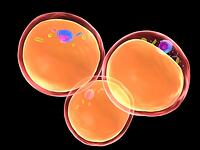
 Inflammatory Events Associated With Obesity
There is an increasing problem with obesity worldwide. In 2008 the World Health Organization reported that 1.5 billion adults (+20yrs) worldwide are overweight (defined by a body mass index of equal to or greater than 25). Of these figures around 500 million were classed as obese with a BMI greater than 30 (1). Obesity has been associated with an increased risk in chronic diseases including cardiovascular disease, diabetes and some cancers. It has also been suggested that some forms of obesity are associated with low grade chronic inflammation. What remains to be elucidated is the primary trigger that is responsible for initiating the inflammatory cascade within adipose tissue. Here, we explore the cell interactions and cytokines associated with adipose tissue and their impact on inflammatory disease.
Adipose tissue - a site of inflammation?
The function of adipose tissue is to store energy in the body for times of calorific restriction but it is apparent that fat is not just simply an inert storage organ. Recent studies have demonstrated a crucial link between fat tissue and inflammation and revealed close interactions between adipocytes and cells of the innate and adaptive immune response. Adipocytes release bio-active molecules known as adipokines, as well as secreting pro-inflammatory cytokines like IL-6, IL-8 and TNF (2). In particular, the key adipokine known as adiponectin is found at a high plasma concentration and can enhance insulin sensitivity as well as having anti-inflammatory properties. Adiponectin deficient mice have increased expression of endothelial adhesion molecules as well as increased leukocyte binding (2).Furthermore, adipocytes release chemoattractants such as monocyte chemotactic protein (MCP-1) that are able to draw monocytes from the blood into adipose tissue. Elevated MCP-1 levels correlate with weight gain and are over-expressed in adipose tissue (3) resulting in increased numbers of adipose tissue macrophages (ATM), a phenomenon which has been demonstrated in rodent models and in humans (4). These adipose resident macrophages shift from a protective alternatively activated (‘M2’) macrophage state to a more classical pro-inflammatory ‘M1’ state due to pro-inflammatory cytokines involved in activation of macrophages like TNF-α and IL-6 (5), a crucial process in inflammation associated with obesity.
ATMs have been well characterized in their involvement in insulin resistance in obese individuals. Using leptin (ob/ob) or leptin receptor (db/db) deficient mice and diet induced obesity (DIO) in C57BL/6 mice Xu et al demonstrated that the expression of genes related to inflammation are restricted to macrophages in the white adipose tissue (WAT). Using PPARγ agonists such as Rosiglitazone, which regulate adipocyte differentiation and are used therapeutically for treating insulin sensitivity, the mRNA levels for inflammation genes such as MIP-1α, MCP-1, ADAM can be down regulated in ob/ob mice. (6). Furthermore, upregulation of genes associated with inflammation were not found to be abundant in muscle, liver or spleen of obese mice. The expression of inflammatory specific genes in adipose tissue macrophages is reported to occur before the increase in insulin resistance. This suggests that inflammation associated with obesity may be crucial in the development of obesity related insulin resistance which can lead to type II diabetes (6). These findings suggest a strong correlation with inflammation and white adipose tissue in obesity. Further to this, there is a strong correlation with macrophages in adipose tissue and insulin resistance in mouse models, demonstrated by evaluating the temporal expression of macrophage marker levels in the WAT of mice fed a high fat diet. It was found that the expression of macrophage markers were up-regulated before the increase in circulating insulin levels at 16 weeks of high fat diet. These results suggest that macrophage activity occurs after the increase in adipose tissue but before the insulin resistance (5). Interestingly, lymphocyte infiltration to white adipose tissue has been shown to precede the activation of macrophages in white adipose tissue (7).
Furthermore, there is compelling evidence to suggest that IL-17 could be a key link between adipose tissue and inflammation. IL-6 production in association with adipose tissue has led investigation into Th-17 differentiation and IL-17 associated autoimmunity in obesity. T-cells from diet induced obese (DIO) mice expand TH-17 cells and produce higher levels of IL-17 than lean controls in an IL-6 dependent manner (8). This bias towards a TH-17 like state may be linked to exacerbation of autoimmune diseases. However, it is unclear how this relates to macrophage activation in white adipose tissue, but it is clear that the process is much more dynamic than involving only a few cell subtypes.
References
- World Health Organization
- Vachharajani, V et al. (2009) Life. 61(4), 424-430
- Rasouli, N. (2008) J Clin. Endocrinol. Metab. 93: s64-s73
- Nishimura s. et al (2009) Inflammation and Regeneration 29, 118-122
- Lumneg, C.N. (2007) The Journal of Clinical Investigation. 117, 178-184
- XU, H et al. (2003) J. Clin. Invest. 112:1821-1830
- Caspar-Baguil. S et al (2009) Journal of physiology and biochemistry. Volume 65, Number 4, 423-436
- Ahmed, M. (2010) Cytokine and Growth Factor Reviews. 449-453
- Wellen, K. E et al (2003) The journal of clinical investigation. 112, 1785-1788


