<img height="1" width="1" style="display:none;" alt="" src="https://dc.ads.linkedin.com/collect/?pid=217082&amp;fmt=gif">

Preclinical Laboratory Services

6OHDA Model of Parkinson's Disease

Evaluating neuroprotective therapies for Parkinson's Disease

6OHDA-induced Parkinson's Disease (PD) Efficacy Model

  • Our 6OHDA (6-hydroxydopamine) induced model is a unilateral lesion model of which the Nigro-Striatial pathway is damaged. Owing to the fact that systemically administered 6-OHDA fails to cross the blood-brain barrier, we instead stereotaxically inject toxin directly into the brain region of interest, typically the SN, ventral tegmental area (VTA) or striatum (5).
  • The size of the resulting lesion depends on the amount of 6-OH- DA and size of bolus administered, and is typically performed unilaterally; bilaterally-injected animals require intense nursing. Moreover, the non-injected hemisphere nicely serves as an internal control.
  • Upon injection, 6-OHDA is preferentially taken up by DAergic neurons through the DA transporter system (DAT), and thus specifically kills DAergic neurons while sparing other cell types.
  • When inside neurons, 6-OHDA exerts is neurotoxic effects by inhibiting complex I of the mitochondrial respiratory chain and participating in the formation of superoxide free radicals.
  • Lesions in the SN-VTA develop rapidly, with neuronal death occurring within ~12 hours. Intriguingly, intrastriatal injections show a much slower progressive degeneration that occurs retrogradely.
  • Beginning several days after unilateral injection of 6-OHDA, the mice exhibit a quantifiable contralateral turning behavior induced by systemic administration of a DA agonist or amphetamines.
  • Since injections into the SN-VTA display acute effects, some preferred uses include development of new pharmacological agents and cell replacement thera- pies. On the other hand, the more prolonged degeneration that occurs with injections into the striatum lends itself nicely to exploring neuroprotective therapies. 

Assessments:

  • Rotations
  • Paw Placement (scroll down to see test results) 
  • Histology/IHC

Don't see the model or assessment you require? Our scientists are experienced with rapidly validating models from the literature. Please speak with a scientist to discuss your requirements. 


Parkinson's Staining 

We also perform specific staining methods for Parkinson's research, including:

IHC staining using Tyrosine Hydroxylase (TH) antibody - used for staining of Dopaminergic neurons, as TH is one of the enzymes involved in the production of Dopamine in neuronal cells. This staining is used in MPTP-induced PD model in mice or 6-OHDA-induced PD model in rats.


Staining of 6-OHDA-induced Parkinson’s Disease Brain Sections in Rats

Screen Shot 2017-02-03 at 1.08.24 PM.png

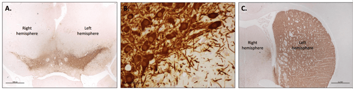
Figure A. Subgross image of Substantia Nigra sections showing reduction in number of TH-positive cells in the right hemisphere vs. the left intact hemisphere. Figure B. High magnification of TH-positive dopaminergic neurons. Figure C. Subgross image of TH-positive nerve fibers in section of striatum of the Left intact hemisphere contrasted to the lack of TH staining in the right hemisphere injected with 6-OHDA.  


SPECIES AVAILABLE:

  • Rat 6OHDA


Request Study Proposal

 Scientists-Smiling-Laboratory-Clinical-Research.jpg

CONTACT US:


+1-651-641-1770

1-888-876-3246

+41-44-986 2628

Contact Us