<img height="1" width="1" style="display:none;" alt="" src="https://dc.ads.linkedin.com/collect/?pid=217082&amp;fmt=gif">

MD Biosciences Blog

IHC Staining Image Gallery Now LIVE: High resolution whole slide image samples from in-house histopathology laboratory

Posted by MD Biosciences on Mar 21, 2019 11:57:33 AM

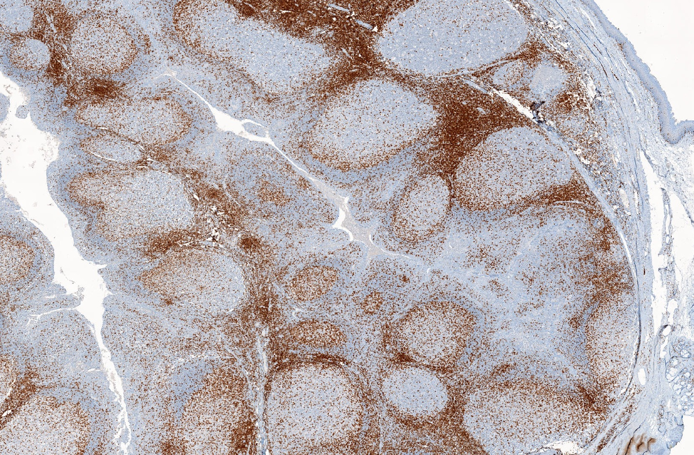
 MD Biosciences has extensive experience in the application of immunohistochemistry to analysis of markers of interest in tissue. Our validated protocols permit such assessments as lymphocyte analysis, cellular proliferation, cellular activation/differentiation, inflammation, biomarker analysis, and much more.

View high-resolution whole slide image examples of our IHC staining results below!

https://www.mdbhistopath.com/immunohistochemistry-image-gallery-ihc-markers 


Don't see a marker you're interested in? We provide custom IHC assay development and validation using either commercially available or Sponsor-provided antibodies, and additionally have in-house antibody development facilities for challenging targets. Contact us to discuss a custom project! 

Contact Us Today!  651-641-1770

Read More

Topics: CRO/outsourcing, Biomarkers, Imaging, Pathology, GLP Histopathology, clinical research, Histology, GLP, In-situ Hybridization, Immunohistochemistry, Tissue Processing

IHC Staining Image Gallery featuring CD3, T-cell co-receptor in tonsil tissue

Posted by MD Biosciences on Mar 15, 2019 12:20:36 PM

Below is a sample staining that demonstrates anti-CD3 IHC, 2x. CD3 is the T-cell co-receptor and is considered a Pan-T Cell lineage marker. T cells are seen surrounding B Cell Follicles in the below tonsillar tissue, demonstrating rich T-cell zones and interspersed t-cells within follicles. More high IHC quality images available upon request, and pathology review available in house.

Call us today for your histology needs: (651) - 451-1770.

Contact Us

ValidationSlides_Tonsil_IHC_CD3_PanTCellMarker_2x-1

Read More

Topics: Histology, GLP, Immunohistochemistry

MD Biosciences GLP-approved Histopathology Laboratory launches new website: featuring histology, IHC, ISH and digital pathology services

Posted by MD Biosciences on Feb 15, 2019 9:00:00 AM

Our new histopathology website is now live! Check it out here!

MD Biosciences Laboratories offer a broad range of histology, immunohistochemistry (IHC) and In-situ hybridization services, staining services, tissue processing and digital pathology to enhance readouts and study applicability to the clinic.

We operate two distinct histology and IHC laboratories, including a GLP developmental research laboratory and a CLIA clinical IHC laboratory, offering a broad range of state-of-the-art technical capabilities to meet research and clinical study needs. Method development and optimization in addition to routine analysis shapes the foundation for our tissue-based services. The laboratories are equipped with advanced automated staining systems and digitized imaging, facilitating the privileged access to large, annotated image files. Services are complimented by the Visiopharm artificial intelligence-based image analysis platform to enhance the quality and speed of analysis. Services are supported by expert consultant pathologists specialized in various areas. 

Contact us to learn more about our services!

Contact Us

Read More

Topics: CRO/outsourcing, Pathology, GLP Histopathology, Histology, GLP, Artificial Intelligence, In-situ Hybridization, CLIA certified, Immunohistochemistry, Tissue Processing